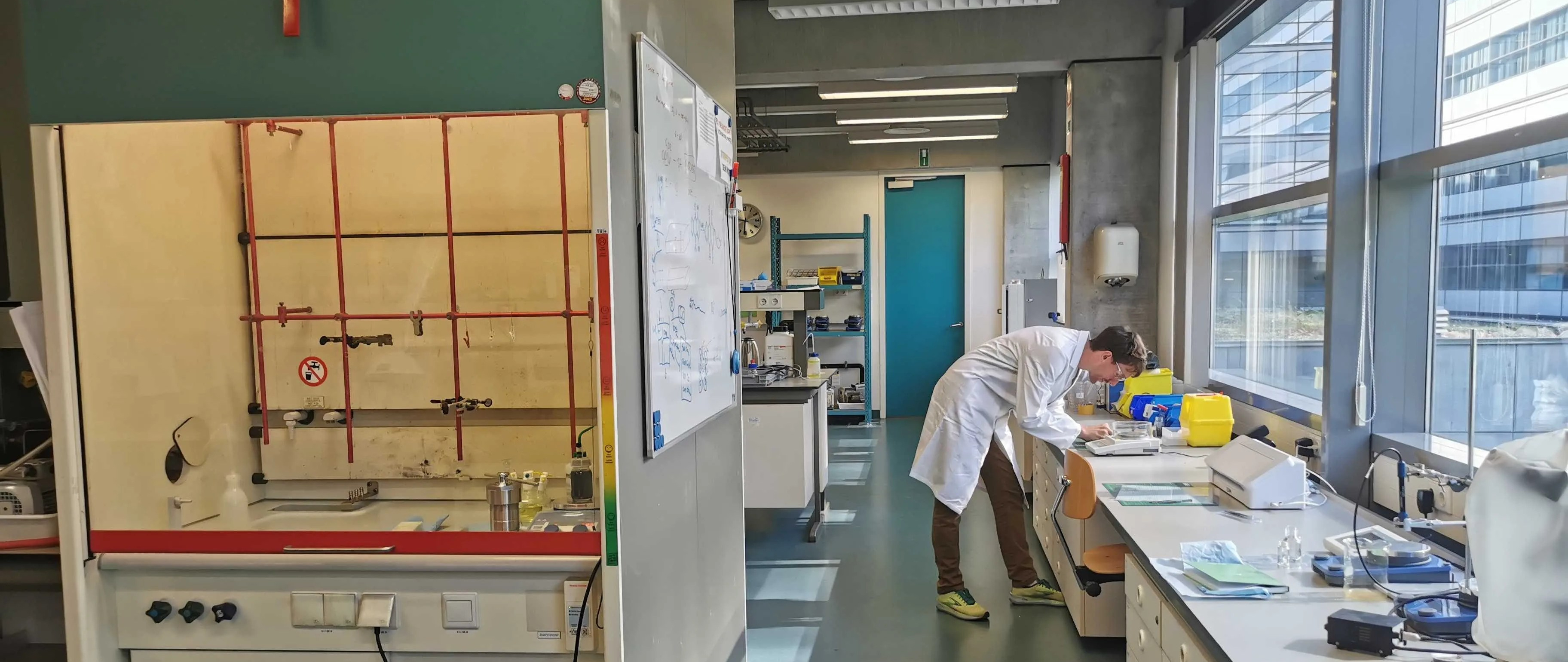
Welcome in the lab!
A glimpse into my lab work and aspects of science I deem curious enough to write about. Feel free to look around and ask questions if anything peaks your interest!
Temporary note: While I am working through a backlog of images, some updates might not be in chronological order.
30/04/23
Happy accident
I often used the analogy to X-rays in the hospital when explaining my work related to X-ray tomographic imaging to people unfamiliar with the topic. Having worked with X-ray tomographic imaging since the start of my PhD on fuel cell materials, it was quite exciting when a bike accident “forced” me to have a CT scan of my wrist taken. This segmentation of the fractured head of my radial bone was definitely one of the most exciting data sets I ever had the chance to work on.
Segmented reconstruction of an X-ray CT scan of my right wrist after a bike accident. Clearly visible is the fracture in the head of the radial bone and maybe also a small crack in the head of the ulna, but I am no medical expert.
CT scanner: *Radiology Máxima MC Veldhoven
Software: (Fiji) ImageJ
ParaView

11/04/23
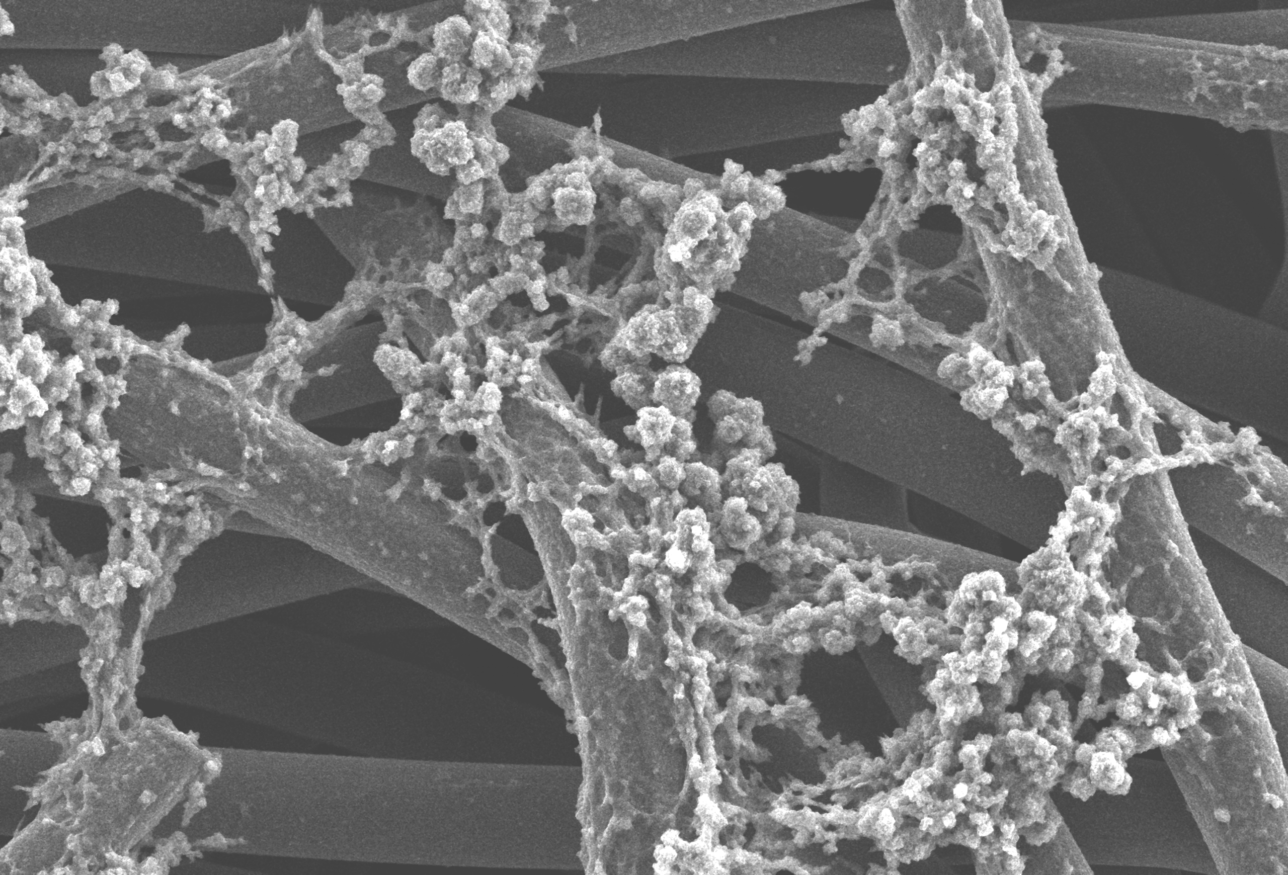
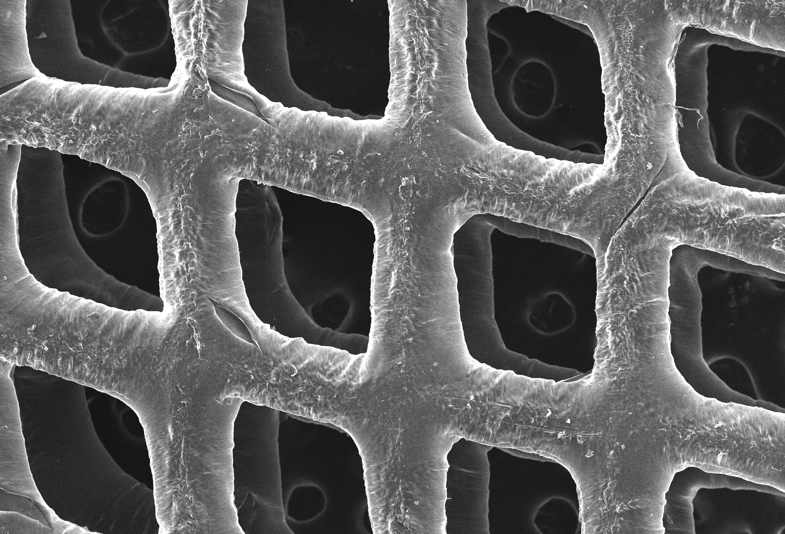
Curiosity
Otherworldly structures, hidden to the naked eye. While we often use analytical devices purely to extract qualitative and quantitative information, I very much enjoy exploring the world hidden in the orders of magnitude below our perception.
A collection of SEM images from the work of some of my colleagues.
SEM: JEOL IT 100
Software: (Fiji) ImageJ


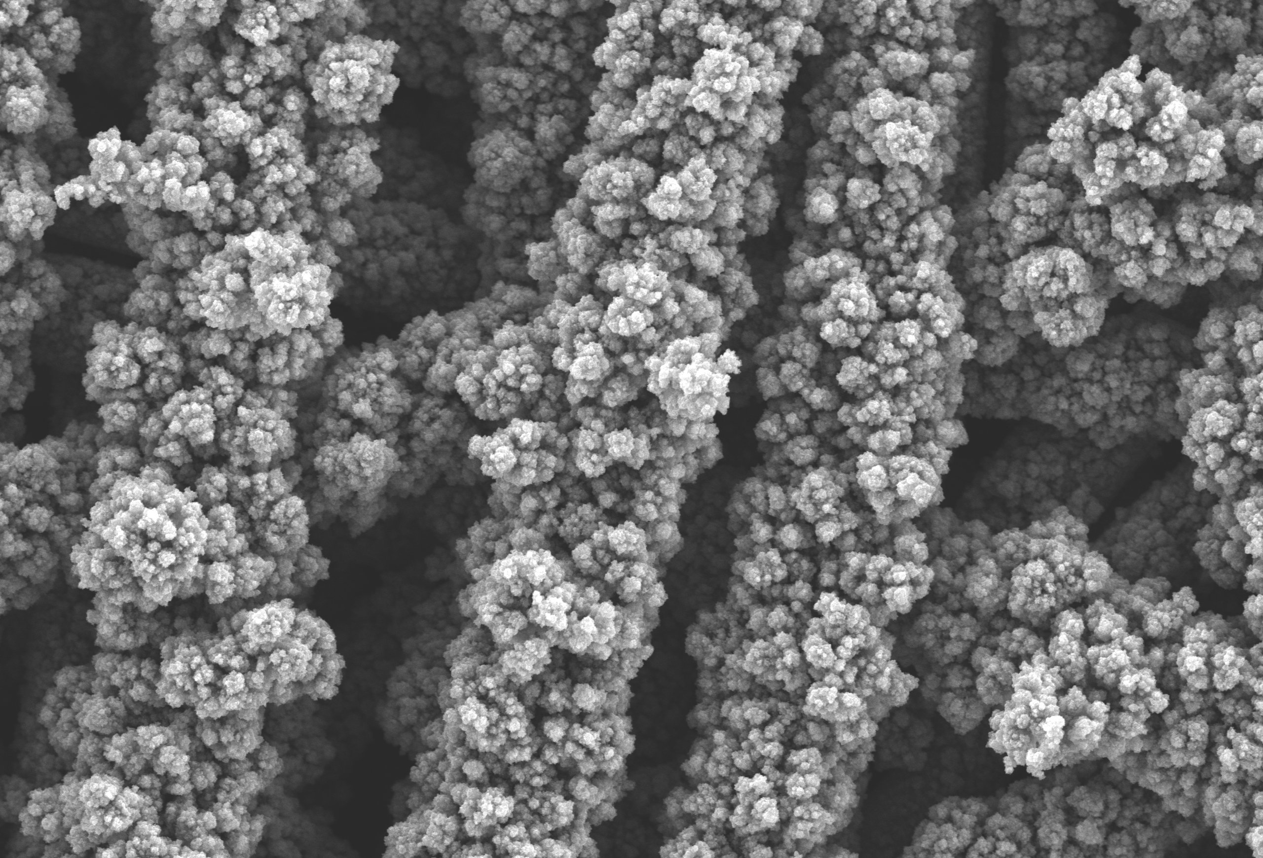

09/11/2021
216
That’s how many minutes it took to acquire the 72 SEM images I used to make this visualization of an early version of a DHBT foam. The feature in the center was an undesired result of non-optimized of parameters during the synthesis but made for a great focal point during imaging and processing.
SEM images obtained at different rotation angles of the sample holder, registered to a common feature.
SEM: JEOL IT 100
Software: (Fiji) ImageJ

